
FynSig: Bringing AI Precision to Biomedical Cell Analysis

A single laboratory instrument can capture thousands of images of human cells every second. But identifying and counting those cells accurately, particularly when they overlap or clump together, remains a significant challenge for biomedical researchers.
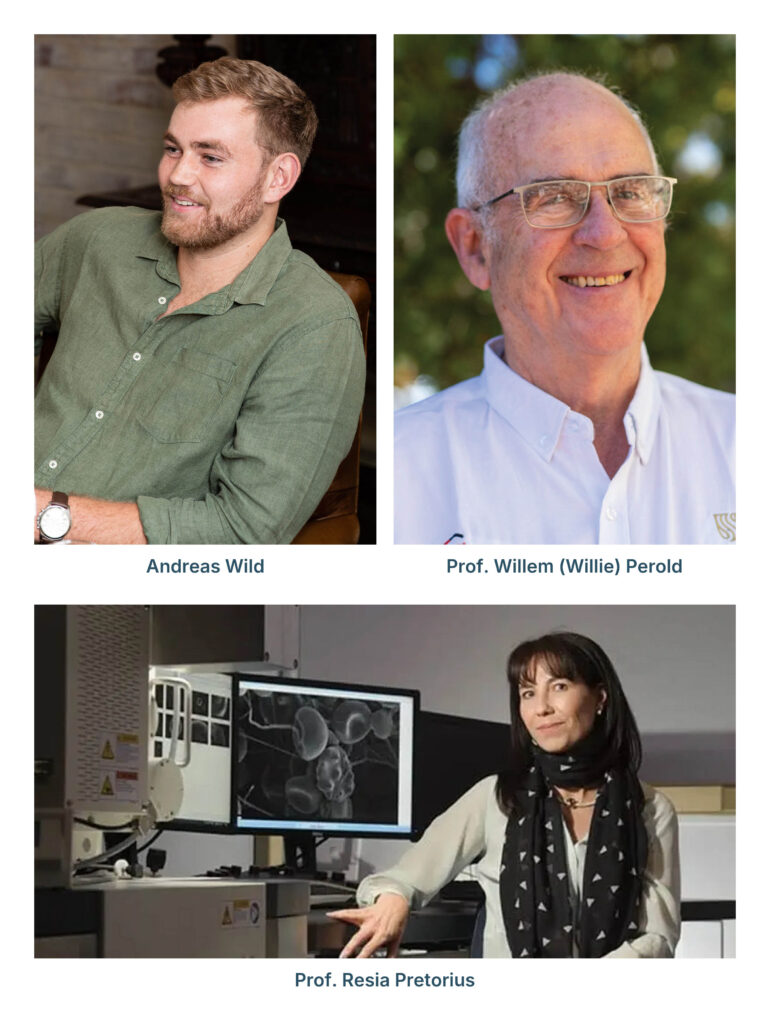
FynSig Biotech, an emerging biomedical technology venture founded by biomedical engineer Andreas Wild, is developing artificial intelligence (AI) technology to make cell analysis faster, more accurate, and accessible, with the ambition of helping clinical trial and pathology laboratories process large volumes of complex data more efficiently.
Wild was among the innovators at Cape HealthTech Connect 2026, held in September at Stellenbosch University's Biomedical Research Institute. His work reflects the research-driven entrepreneurship championed by SWEAT Africa, demonstrating how AI can address practical challenges in biomedical research.
The venture emerged from a challenge Wild encountered while working with the blood research group led by Professor Resia Pretorius at Stellenbosch University in early 2025.
"I always knew that biology is extremely complex, but I got new respect when I spent some time in an actual lab during my postgraduate studies," he explains.
The group was investigating changes in blood associated with severe illness, including the tendency of cells to stick together. Existing analysis methods struggled to distinguish individual cells within these clusters.
For Wild, whose expertise lies in computer vision, the problem presented an opportunity. "I thought: 'This is a problem I know how to solve!'"
The technology centres on imaging flow cytometry, which combines rapid cell analysis with microscopic imaging. As cells flow through a narrow channel, a specialised microscope captures approximately 1 000 images per second. Researchers analyse these images to identify and count cells, generating information relevant to disease research and immune responses. CD4 white blood cell counting, for example, is an important measure of immune function in HIV care.
However, when cells overlap or clump together, conventional image analysis can mistakenly count them as a single object.
"Simply put, our technology counts each individual cell as its own object instead of merging them together as one big blob," Wild explains.
This capability is particularly relevant to inflammatory disease research, where cell aggregation may itself be biologically significant.
Although powerful computer vision models already exist, many are too large or computationally demanding for imaging flow cytometry and struggle with the low-resolution, motion-blurred images these instruments produce.
FynSig's solution is a lightweight AI model of approximately 1.3 MB that can run locally on ordinary laptops, avoiding the need to transfer sensitive patient data to external servers. According to Wild, benchmarking on the team's dataset demonstrated accuracy comparable to existing state-of-the-art models, with faster processing speeds.
The model has been trained and tested in academic work awaiting peer review. An interactive analysis platform is now being developed to allow researchers to visualise and investigate their data without writing code.
A defining feature is the emphasis on explainable AI, allowing researchers to understand and interpret the technology's results.
"I am a massive fan of explainable AI, even more so in the biomedical setting," Wild says. "This project is about keeping humans in the loop and speeding up their analysis."
FynSig also aims to support conventional flow cytometry workflows, making its technology useful to laboratories that have not upgraded to newer imaging instruments.
Turning the research into a practical product has reinforced another lesson: the importance of listening to potential users. Wild credits Stellenbosch University's LaunchLab, recalling advice from its head, Brandon Paschal, to "talk to humans".
"Talking to humans and hearing their feedback is the most important thing to get started," he says. FynSig plans to work with early adopters to refine the platform around their needs.
The name FynSig, meaning ‘fine-sighted’ in Afrikaans, was inspired by Wild's walks through Stellenbosch's Botmaskop Nature Reserve, where many of his ideas took shape.
Looking ahead, Wild hopes the technology will reduce laboratory analysis times and costs. Feedback from laboratories suggests that data analysis can take twice as long as the laboratory work and data acquisition themselves.
Beyond efficiency, more detailed cell analysis could help researchers identify biological patterns that existing methods miss, potentially supporting inflammatory disease research, drug discovery, and clinical trials.
Ultimately, FynSig aims not to replace scientific expertise, but to equip researchers with faster, more accessible tools for understanding complex biological data, while keeping human judgement at the centre of the process.
For more info: www.fynsig.com
News date: 2026-10-30
Links:
KRISP has been created by the coordinated effort of the University of KwaZulu-Natal (UKZN), the Technology Innovation Agency (TIA) and the South African Medical Research Countil (SAMRC).


Location: K-RITH Tower Building
Nelson R Mandela School of Medicine, UKZN
719 Umbilo Road, Durban, South Africa.
Director: Prof. Tulio de Oliveira